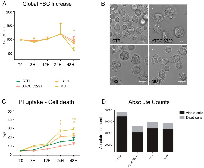
Figure 1.
Morphological features and cell death rate. (A) Trends of forward light scatter (FSC) values for each treatment during the time course from the start (T0) to 48 h. The FSC values were converted to arbitrary units (A.U.), setting the control (T0) to 100. Each value is expressed as the mean ± SD (results from n ≥ 3 independent experiments). Two-way ANOVA with Bonferroni’s multiple comparison test revealed * p < 0.05 vs. control (T0); (B) Bright-field images of monocytes after 24 h of preincubation with Campylobacter jejuni ATCC 33291 lysate (b), C. jejuni ISS 1 lysate (c) and C. jejuni 11168H cdtA mutant lysate (d), compared to the untreated control cells (a). Bars: 10 µm; (C) Trends of percentage of propidium iodide (PI)-positive cells for each experimental condition during the time course from T0 to 48 h. Each value is expressed as the mean ± SD (results from n ≥ 3 independent experiments); * p < 0.05 and ** p < 0.01 vs. control (T0); (D) Absolute counts of viable and dead cells after 48 h of preincubation with the lysates, compared to untreated control cells. Results from a representative experiment.

