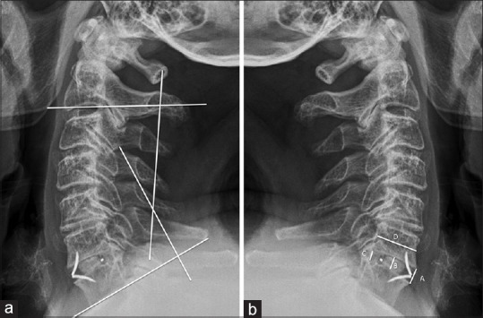
Figure 1.

Lateral radiograph depicting a 4-year long follow-up in a C6“C7 case. (a), Image showing cervical alignment calculated by the Cobb angle between the inferior margins of C2 and C7 vertebral bodies; (b), lateral radiograph showing the disc height measurement. A, is anterior disc height, b is middle disc height, c is posterior disc height, and d is sagittal diameter of the up vertebral body. Disc height index = ([a + b + c]/3)
