Figure 8.

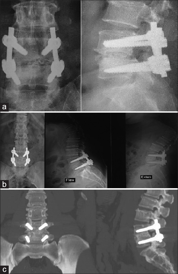
Case Presentation 2: (a) Immediate postoperative X-ray. (b) one year follow up X-ray showing full fusion. (c) One year Computed tomography showing grade 5 fusion

Case Presentation 2: (a) Immediate postoperative X-ray. (b) one year follow up X-ray showing full fusion. (c) One year Computed tomography showing grade 5 fusion