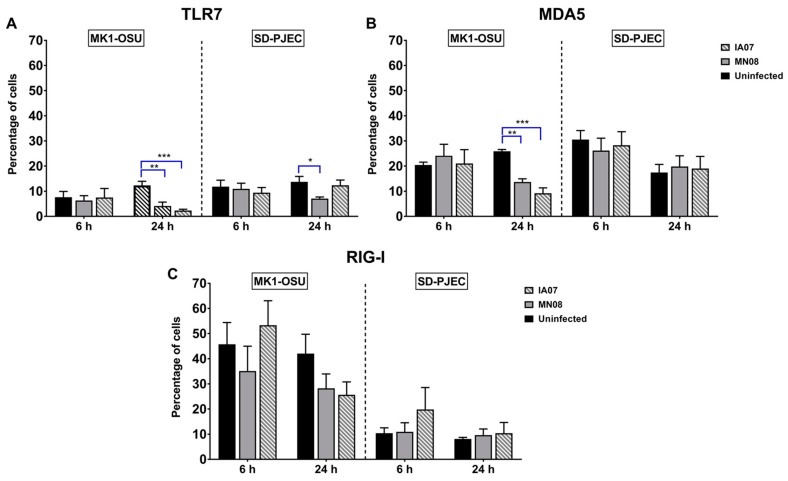
Figure 5.
Expressions of TLRs and RLRs in MK1-OSU and SD-PJEC cells at 6 h and 24 h post-infection determined using a flow cytometer. MK1-OSU and SD-PJEC cells were infected with MN08 and IA07 viruses at MOI of 0.01 and incubated for 6 and 24 h at 37 °C. Cells were fixed and permeabilized using BD Cytofix/Cytoperm, incubated with primary antibodies, treated with biotinylated secondary antibody, and stained with streptavidin-FITC. (A) TLR-7 expression was decreased by both MN08 and IA07 in MK1-OSU cells 24 h post-infection but only by MN08 in SD-PJEC cells. (B) Expressions of MDA5 were reduced by MN08 and IA07 infection in MK1-OSU cells at 24 h but did not affect the expressions in SD-PJEC cells. (C) RIG-I did not alter due to treatments in both cell lines. Bars represent the mean for four to six experiments ± SE p < 0.05 (*) represented a significant difference between uninfected and infected cells. ** and *** represents p < 0.005 and p < 0.0005 respectively.

