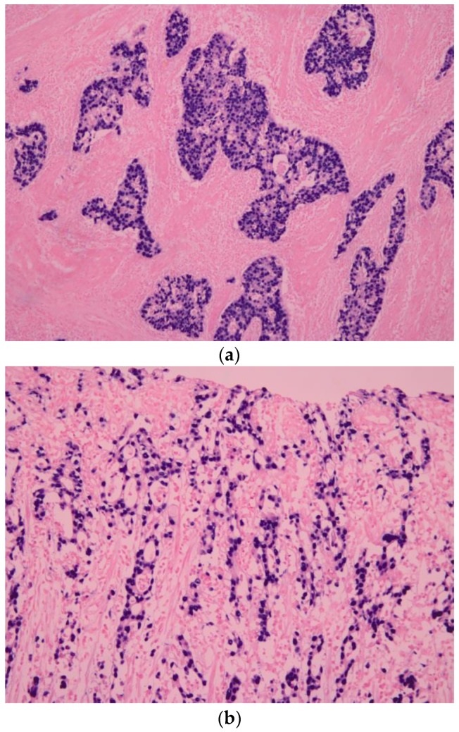
Figure 1.
Histologic characteristic of Epstein–Barr virus-associated gastric carcinoma (EBVaGC). (a) EBV-encoded small ribonucleic acid (EBER1) in situ hybridization shows positive nuclei in the carcinoma cells, which are surrounded by infiltrating lymphocytes (×100); (b) Histologic characteristic of EBVaGC. The “lacy pattern” is composed of irregularly anastomosing tubules and moderate to dense lymphocytic infiltration (×100).

