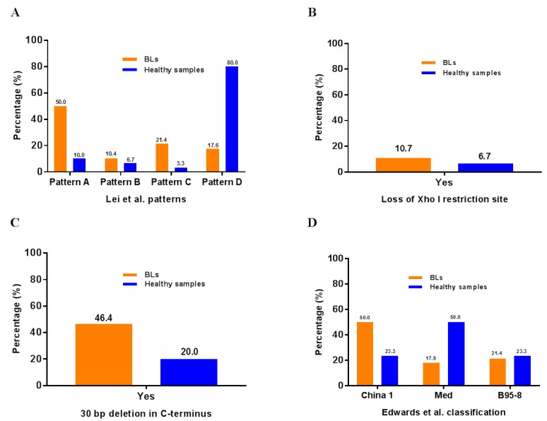
Figure 3.
Different LMP-1 classifications applied to BL and healthy samples analyzed in the study of Lei et al. (2015) [18]. (A) The Patten A–D of Lei et al. [18] study. (B) The loss of Xho I site used in the study of Hu et al. [23] (C) 30 bp deletion in LMP-1 C terminus used in the study of Miller et al. [24] (D) The classification of the LMP-1 C-terminal variants used in the study of Edwards et al. [25].

