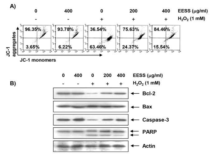
Figure 3.
Attenuation of H2O2-induced mitochondrial dysfunction and changes of apoptosis-related proteins by EESS in SW1353 chondrocytes. Cells were pretreated with the indicated concentrations of EESS for 1 h, and then stimulated with or without 1 mM H2O2 for 24 h. (A) The cells were collected and incubated with 10 µM JC-1 for 20 min at 37 °C in the dark. The cells were then washed once with PBS, and the values of MMP were evaluated by flow cytometry. The data are the means of the two different experiments; (B) The cellular proteins were separated by SDS-polyacrylamide gel electrophoresis, and then transferred to membranes. The membranes were probed with the indicated antibodies. Proteins were visualized using an ECL detection system. Actin was used as an internal control.

