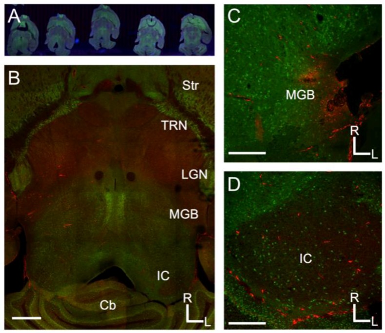
Figure 1.
Projections from the inferior colliculus to the medial geniculate body in the mouse. (A) Overview of Nanozoomer scanned images from serial sections of the mouse brain sectioned to preserve the tectothalamic pathway (see Methods and Lee and Sherman, 2009); (B) Confocal image of a tectothalamic section showing the inferior colliculus (IC) and medial geniculate body (MGB). Other structures visible include the striatum (Str), thalamic reticular nucleus (TRN), lateral geniculate nucleus (LGN) and the cerebellum (Cb); (C) Example of injection site for CTβ-Alexa 594 in the lateral part of the MGB; (D) Labeling in the ipsilateral IC following the injection depicted in C. In each image, green label is the Venus-fluorescent protein and red-label is the CTβ-Alexa 594. Scale bars are 1 mm (B), 500 μm (C), 500 μm (D).

