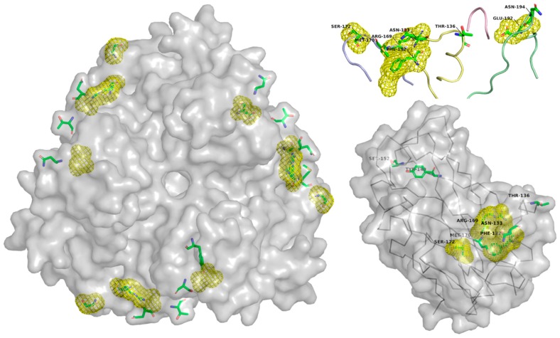
Figure 4.
Membrane cofactor (CD46) interacting residues, and known mutation sites, within the Ad35 fiber knob domain: The key residues which interact with CD46 are shown as green sticks. The yellow surface shows the region in which known mutations which abrogate CD46 interaction occur. In the top right is a detailed view of the 4 loops which interact with CD46, HI (blue), DG (yellow), GH (red), and IJ (green). Structure from PDB: 2QLK.

