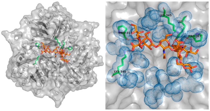
Figure 6.
GD1a/Sialic Acid interacting residues within the Ad37 fiber knob domain. Key residues forming the GD1a-Ad37Fkn interaction are shown as green sticks, with the GD1a in orange, hydrogen bonds are shown by red dashes. While the interface can occur in three orientations, only one set of interacting residues is shown. The blue dots show the surface of all residues shown to be able to interact with GD1a or support the interaction, seen to create a large apical binding pocket. Structure from PDB: 3N0I.

