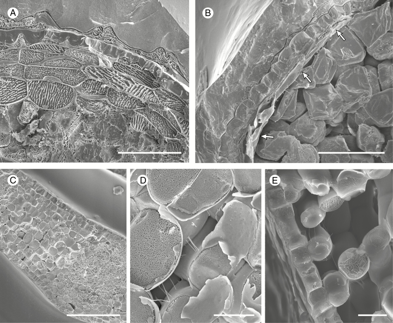
Fig. 2.
Cryo-scanning electron micrographs of Notothylas levieri (A, B) and Hedera helix (C–E). (A, B) Cryo-fractured sporophytes of the estomate hornwort species Notothylas levieri, young (A) and mature (B); note the complete absence of intercellular spaces in the assimilatory layers (A) which collapse and dry out at maturity (B, arrowed). (C–E) Cryo-fractured young leaves. (C) General aspect of a leaf approximately one-tenth its final size with nascent intercellular spaces. (D) Detail of an intercellular space from (C). (E) Spongy mesophyll and lower epidermis in a leaf approximately a quarter of its final size. Scale bars: (C) 100 µm; (A, B) 50 µm; (E) 20 µm; (D) 10 µm.

