Figure 4.
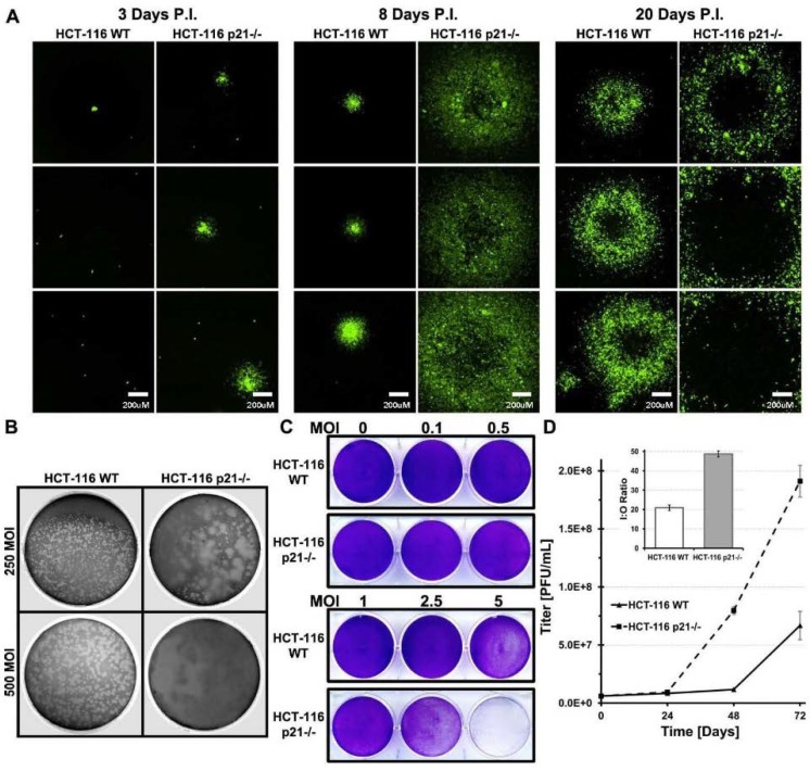
Viral burst size is significantly larger in HCT-116 p21−/− cells. HCT-116 WT and p21−/− cells were plated in 10-cm dishes and infected with 250 or 500 PFU of AdTrack-HisE1A-E1B virus. After 1 h, virus was removed and overlaid with 0.1% Agarose Media mixture. GFP fluorescence was imaged at indicated time points to follow viral burst (A). HCT-116 WT and p21−/− cells were plated in 10-cm dishes and infected with 250 or 500 PFU of CN702 virus. After 1 h, virus was removed and overlaid with 0.1% Agarose Media mixture. 26 days p.i. overlays were stained with neutral red overnight (B). HCT-116 WT and p21−/− cells were infected with various MOI of CN702. At 72 h, p.i. cell monolayers were stained with 0.5% crystal violet in methanol to visualize viral CPE (C). Assessing infectious particle titer over time. HCT-116 WT and p21−/− cells were plated in 6 well plates and infected with 10 MOI of CN702. Cells and media were harvested at indicated time points and viral titer was done on 293 HEK cells (D). Statistical significance was defined as * p ≤ 0.05.

