Figure 1.
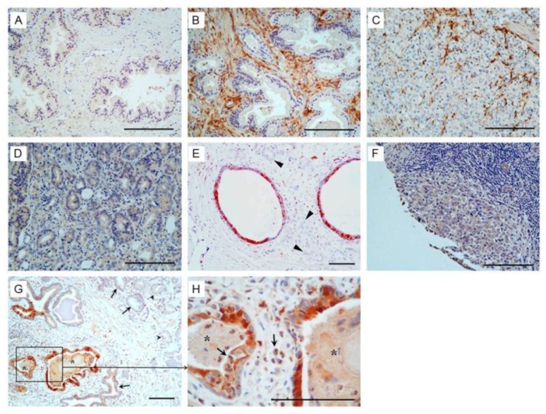
Immunohistochemistry analysis of Prostate-associated Gene 4 (PAGE4) in prostate cancer. (A) Negative staining in the normal prostate. (B) Intense staining shown in the stromal tissue in benign prostatic hyperplasia (BPH). (C) Positive staining in the stromal cells but negative in the cancer cells in some prostate cancer (PCa) specimens. (D) Moderate staining in the cancer cells but negative in the stromal cells in some PCa specimens. (E) Positive staining in the atrophic glands but negative in the cancer cells (arrowhead). (F) Negative staining in metastatic PCa. (G) Intense staining shown in cancer adjacent “normal” glands (asterisk) associated with inflammation but only moderate staining in the cancer cells (arrowhead). (H) High power view of boxed area in (G). Asterisk, proliferative inflammatory atrophy (PIA) lesions; arrows, inflammatory cells. Scale bars in all panels, 100 μm.

