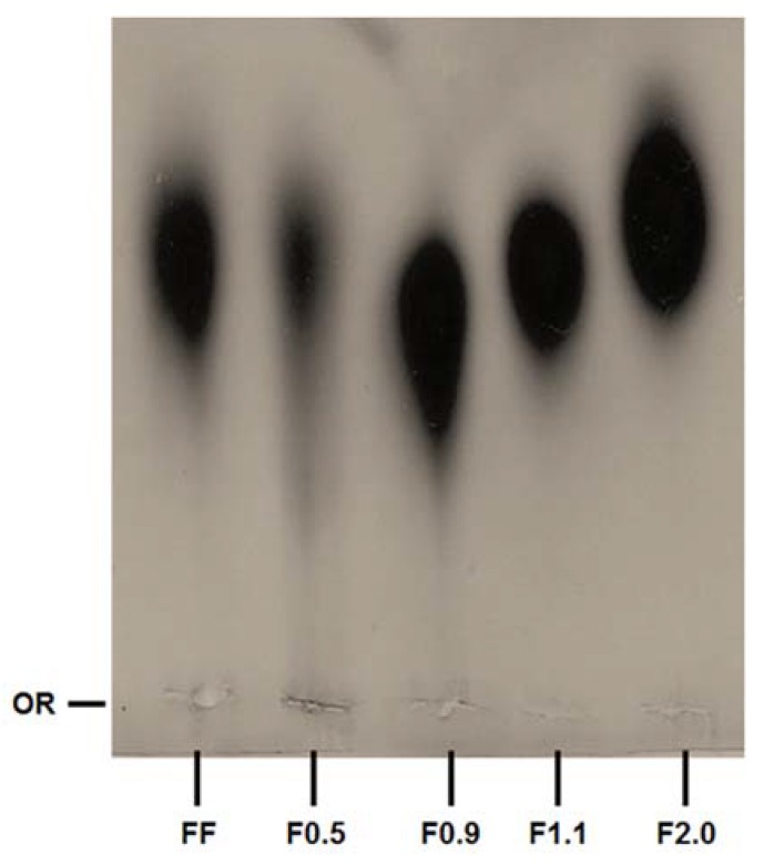
Figure 1.
Staining pattern of the polysaccharides after agarose gel electrophoresis, stained with toluidine blue. About 5 µL (50 µg) of each sample was applied in agarose gel prepared in diaminopropane acetate buffer and subjected to electrophoresis, as described in methods. OR—origin. This figure is representative of three separate tests made independently.

