Figure 2.
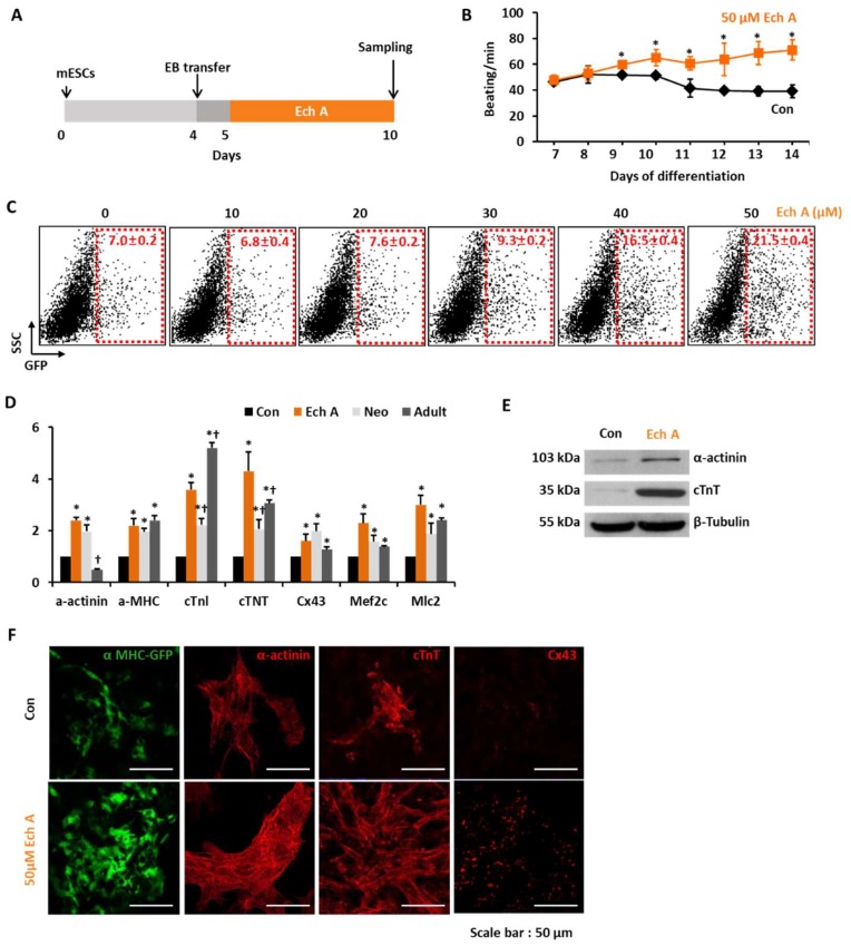
Ech A enhances cardiomyocyte differentiation from mouse embryonic stem cells (mESCs). (A) The protocol for the differentiation of cardiomyocytes from the mESCs using the hanging drop method to produce differentiated embryoid bodies (EBs). (B) The beating number per minute (BPM) of EBs during differentiation. Each group, n = 3. * p < 0.05 (C) Representative FACS analyses and the percentage of mESC-derived αMHC+ cardiomyocyte-like cells according to the dose of Ech A. Each group, n = 3. (D) Relative mRNA expression levels of cardiomyocyte specific genes in nontreated control (Con), Ech A treatment (Ech), heart tissue from neonatal (7 day, Neo) or adult mice (10 weeks, Adult). Each group, n = 3. * p < 0.05 vs. Con, †p < 0.05 vs. Ech A. The mRNA level of glyceraldehyde-3-phosphate dehydrogenase (GAPDH) was used as an internal control. (E) Western blot analyses for the expression of cardiomyocyte specific proteins incubated with the control vehicle (Con) or 50 µM of Ech A. (F) Images displaying αMHC-GFP+, α-actinin+, cTnT+, and Cx43+ cells incubated with Con or 50 µM of Ech A (scale bars, 50 μm). SCC: side scatter cell, cTnI: cardiac troponin I, cTNT: cardiac troponin T, Cx43: connexin 43, Mef2c: Myocyte Enhancer Factor 2C, Mlc2: Myosin light chain 2.

