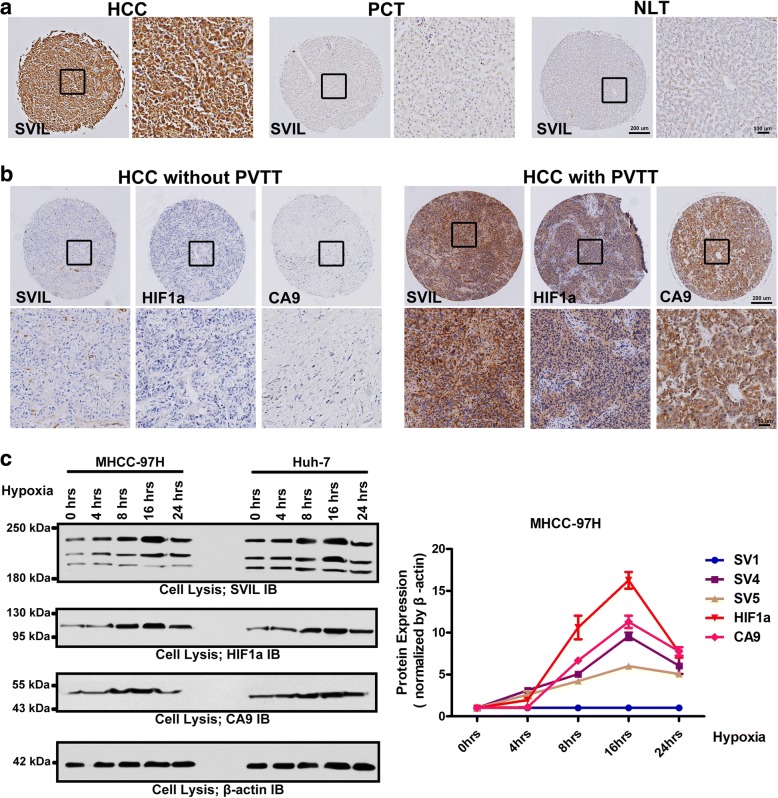
Fig. 1.
Supervillin is positively associated with portal vein tumor thrombus (PVTT) development and metastasis in hepatocellular carcinoma (HCC) patients. a. Immunohistochemical (IHC) staining showing upregulation of supervillin in HCC, compared with para-carcinoma tissue (PCT), and normal liver tissue (NLT). The left image is under 40× magnification and the right image (inset box) is under 100× magnification (black). b. IHC staining showing the accumulation of HIF1α, CA9 and supervillin in a tumor with PVTT, compared with a tumor without PVTT. The upper image is under 40× magnification and the lower image (inset box) is under 100× magnification (black). c. Western blot analysis showing the expression of supervillin, HIF1α and CA9 in MHCC-97H and Huh-7 cells. β-actin was used as the loading control. Data represent the mean of at least three independent experiments ± SD in MHCC-97H cells

