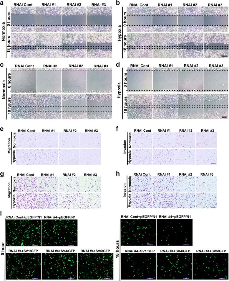
Fig. 2.
Supervillin promotes HCC migration and invasion during hypoxia. a-d. Huh-7 (a, b) and MHCC-97H (c, d) cell mobility was detected by wound healing assay. Cells were transfected with control or supervillin-specific siRNA and incubated under normoxic conditions for 48 h, scratched and exposed to normoxia, or hypoxia for 18 h, respectively. The closure of the scratch was monitored and photographed. Scale bar = 200 μm. E, G. Huh-7 (e) and MHCC-97H (g) cell migration were detected by Boyden Chamber Transwell assays. Cells were transfected with control or supervillin-specific siRNA and incubated under normoxic conditions for 48 h, after which they were seeded into Transwell chambers for 18 h under normoxia or hypoxia. The number of migrated cells was monitored and photographed. f, h. Huh-7 (F) and MHCC-97H (H) cell invasion were detected by Boyden Chamber Transwell assays. Cells were transfected with control or supervillin-specific siRNA and incubated under normoxic conditions for 48 h, seeded into Matrigel-coated Transwell inserts, and incubated under normoxia or hypoxia for 18 h. The number of invaded cells was monitored and photographed. i. MHCC-97H cells that had been treated with supervillin-specific siRNA (RNAi #4, targeted for the supervillin 3′UTR) were allowed to recover their expression of SV1, SV4, and SV5 before assay for cell migration in Transwell chambers under hypoxia for 18 h. The migrated cells on the upper side (at 0 h) and lower side (18 h) were monitored and photographed

