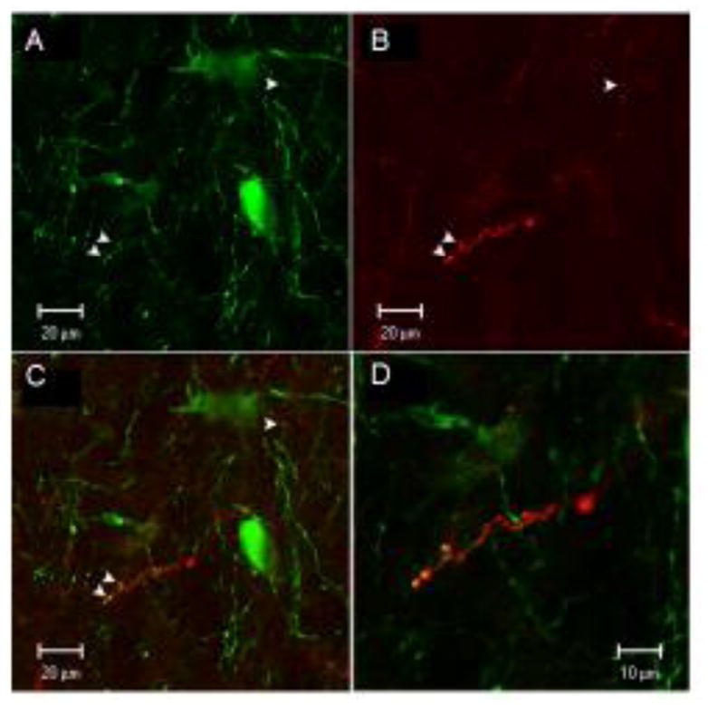
Figure 4.

Confocal images showing neuron profiles labeled with green TH-ir in the A7 cell group (A), axons labeled with Fluoro-Ruby from the injection site in 1B (B), and co-localization of Fluoro-Ruby labeled axons with TH-ir dendrites (C). Arrows show yellow puncta at sites of co-localization. Panel D is an enlargement of panel C.
