
Figure 5.
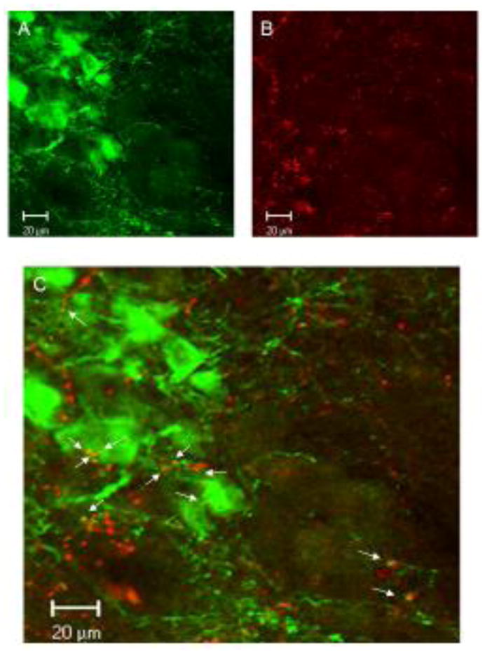
Confocal images of TH-ir neuron profiles in the A7 area (A) with Fluoro-Ruby labeled axons form an injection in the LH similar to that shown in 1B (B), with co-localization of the axons with TH-ir dendrites and cell bodies, as shown by yellow puncta (arrows; C).