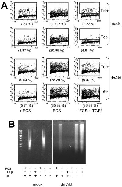
Figure 7.
Expression of dominant-negative Akt blocks the antiapoptotic effect of TGFβ1. (A) Control (mock) and dn Akt HaCaT keratinocytes were incubated in serum-containing or serum-free medium ± Tet for 72 h in the presence or absence of 2 ng/ml TGFβ. The proportion of apoptotic cells was assessed by Apo-BrdU analysis and is indicated in parentheses. (B) Mock and dn Akt HaCaT cells (106 cells/dish) on 60-mm dishes were incubated in medium containing FCS or in serum-free medium ± 2 ng/ml TGFβ for 72 h. To suppress dn Akt, Tet was added during the incubation period where indicated. Adherent and floating cells were pooled, their DNA was collected, and next evaluated for evidence of internucleosomal fragmentation in 1.5% agarose gels as described in MATERIALS AND METHODS.

