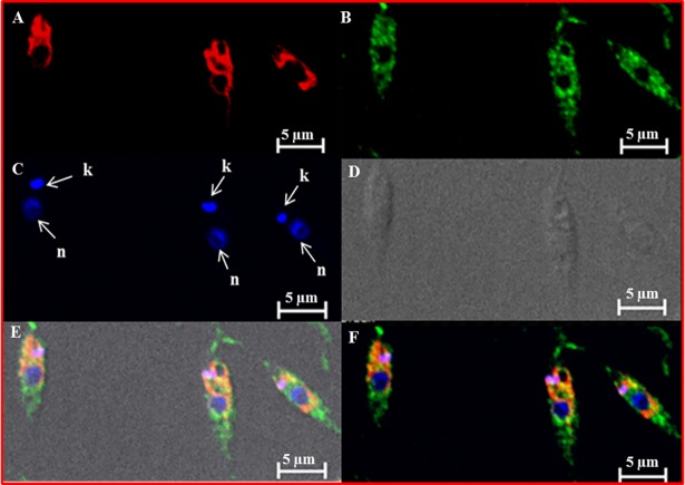
Fig 2. Localization of LdThrRS in L. donovani.
Immunofluorescence analysis by confocal micrograph of wild-type log phase promastigotes stained with DAPI (C), anti-LdThrRS antibody detected using Alexa 488 (green)-conjugated secondary antibody (B) and mitotracker red CMXRos (A). (E) and (F) merged micrographs and (D) phase contrast image. ‘k’ and ‘n’ indicate kinetoplastid and nuclear DNA respectively. The scale bar represents 5 μm.

