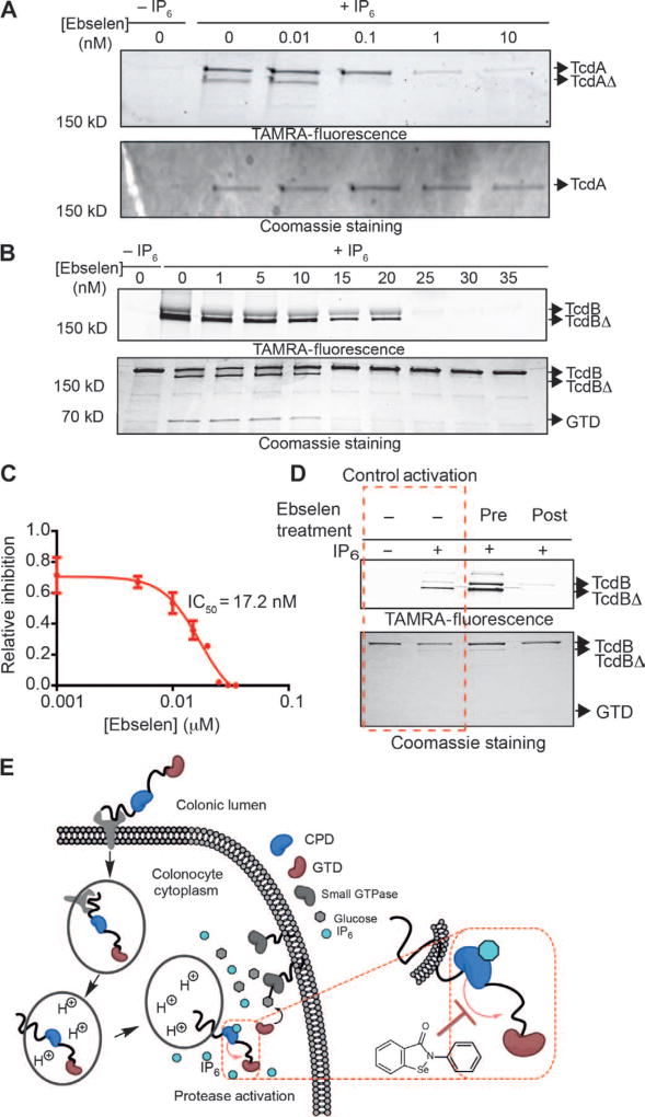
Fig. 3. Inhibition of full-length TcdA and TcdB by ebselen.
(A) TcdA was incubated with increasing amounts of ebselen, activated with IP6, and labeled with TAMRA–AWP-19. Samples were resolved by SDS-PAGE and scanned for probe fluorescence (top gel) or stained by Coomassie (bottom gel). Full-length TcdA and TcdA that has proteolytically released the GTD (TcdAΔ) are indicated by arrows. (B) TcdB was treated as in (A). Samples were resolved by SDS-PAGE and scanned for probe fluorescence (top gel) or stained by Coomassie (bottom gel). Full-length TcdB, TcdB that has proteolytically released the GTD (TcdBΔ), and the free GTD are indicated by arrows. The image shows a typical representation of three replicates. (C) Dose-response curve of ebselen inhibition of TcdB as measured by quantification of probe labeling as shown in (B). Values are plotted for percent inhibition relative to DMSO control (lane 0). (D) Uncoupling ebselen inhibition and IP6 activation. Full-length TcdB was incubated with ebselen before (third lane) or after (fourth lane) size exclusion chromatography. Toxin was then activated with IP6, labeled with TAMRA–AWP-19, and analyzed by SDS-PAGE and in-gel fluorescence analysis. Control lanes without ebselen treatment are marked in the red dashed box. All inhibition experiments were performed at least three times and, where applicable, represented as means ± SEM of triplicate analysis. (E) Proposed point of CPD inhibition mediated by ebselen (red dashed rectangle). Ebselen modifies the active-site cysteine upon IP6-mediated allosteric activation in the cytosol.

