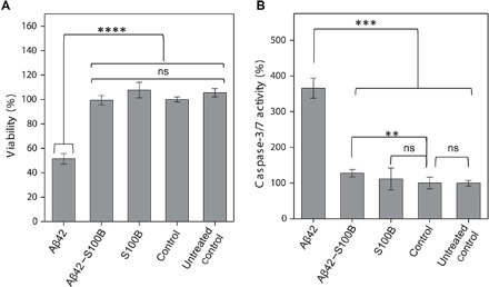
Fig. 5. S100B protects SH-SY5Y cells against Aβ42 toxicity and apoptosis.

(A) Cell viability as measured by PrestoBlue reagent after 72 hours in differentiated SH-SY5Y cells for medium; buffer: 50 mM Hepes (pH 7.4) and 1.1 mM CaCl2, 7 μM Aβ42 with 1.1 mM CaCl2, and 7 μM Aβ42 + 84 μM S100B with 1.1 mM CaCl2. (B) Cell apoptosis as measured by caspase-3/7 activity after 72 hours in differentiated SH-SY5Y cells for medium; buffer: 50 mM Hepes (pH 7.4) and 1.1 mM CaCl2, 7 μM Aβ42 with 1.1 mM CaCl2, and 7 μM Aβ42 + 84 μM S100B with 1.1 mM CaCl2. Results represent mean and SD of two independent experiments (n = 8 and 5 wells per replicates per condition and plate). Statistically significant differences at the 95.0% confidence level using one-way analysis of variance (ANOVA) followed by Welch’s t test. ns, not significant; **P < 0.01, ***P < 0.001, ****P < 0.0001.
