. 2018 Jun 29;7:e33506. doi: 10.7554/eLife.33506

Figure 2. NFRe maintains a low, epidermal expression during root nodule symbiosis.

(A) Transversal root section of nfr1-1-Nin:GUS plants expressing p35S-Nfre shows activation of Nin promoter in the outer cell layer after M. loti inoculation. (B) Transversal root section of nfr1-1-Nin:GUS plants expressing p35S-Nfr1 shows activation of Nin promoter in all cell layers after M. loti inoculation. (C) The epidermal cells, primarily localized in the root susceptible zone, show activity of the Nfre promoter visualized by the nuclear localized triple YFP protein (tYFPnls). (D) Widespread activity of the Nfr1 promoter in the uninoculated root visualized by nuclear localized triple YFP protein (tYFPnls). (E) The expression of Nfre in the susceptible zone of the root, and in the root hairs is maintained after inoculation with M. loti (F). Scale bars, 40 μm (A, B), 0.5 mm (C, D), and 50 μm (E, F).

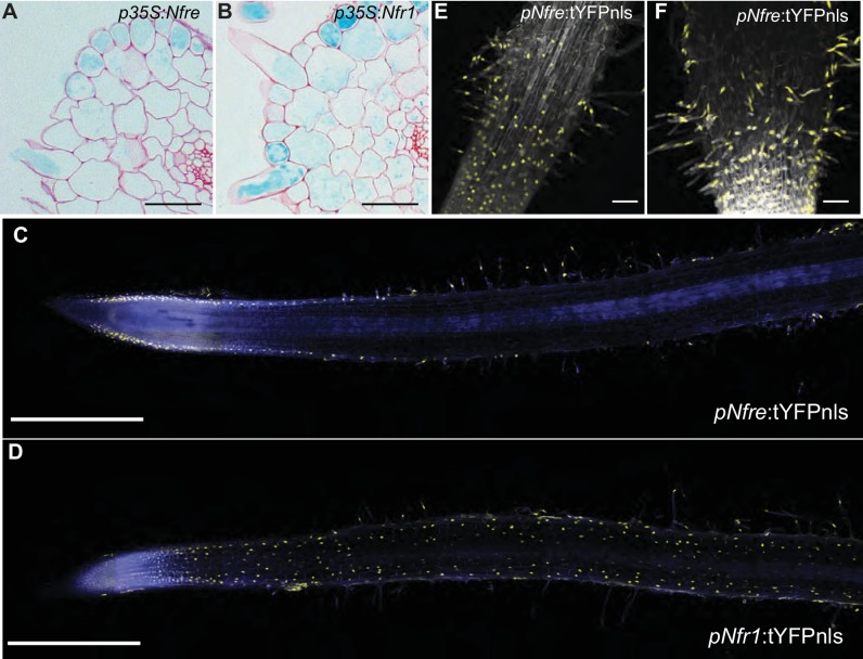
Figure 2.

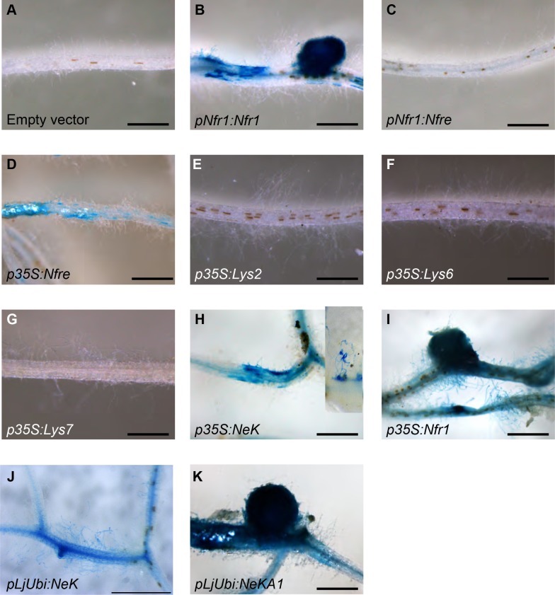
Figure 2—figure supplement 1. Nin:GUS activation in nfr1-1-Nin:GUS plants expressing different receptor variants.

Figure 2—figure supplement 1.

No activation of Nin:GUS was detected in roots transformed with the empty vector (A) containing the endogenous Nfre, when Nfre expression was controlled by Nfr1 promoter (C) or when Lys2 (E), Cerk6 (F), or Lys7 (G) were expressed from 35S promoter. Nin activation but no nodule formation was observed when full length Nfre (D), or chimeric receptor NeK (extracellular region of NFR1, transmembrane and intracellular region of NFRe) was expressed from the 35S (H) or LjUbiquitin (J) promoters. The inset in (H) shows a close-up of observed epidermal Nin:GUS induction. Nodule formation and cortical activation of Nin:GUS was observed in roots expressing Nfr1 under its own (B) or 35S (I) promoter and in roots expressing the chimeric NeKA1 receptor (K) where the activation segment of NFRe was replaced by the corresponding region from NFR1. Scale bars, 1 mm.
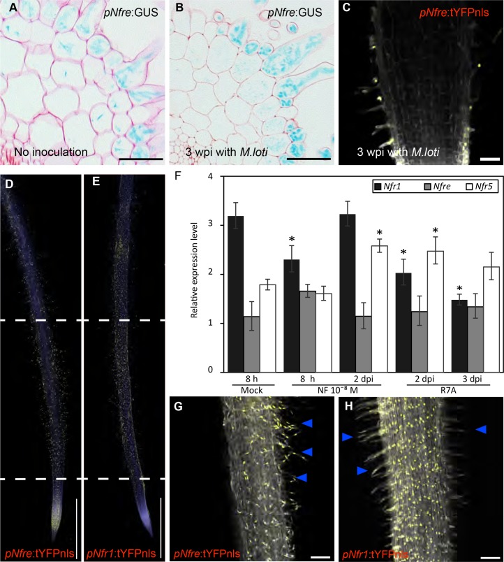
Figure 2—figure supplement 2. Expression patterns of LysM receptors in Lotus japonicus.

Figure 2—figure supplement 2.

(A) The expression of Nfre:GUS in hairy root of WT without Rhizobium inoculation. (B) The expression of pNfre:GUS in hairy root of WT at 3 weeks after inoculation with M.loti (C) The expression of pNfre:tYFPnls is maintained in the root hairs at 3 weeks after inoculation with M.loti. (D) The epidermal cells, primarily localized in the root susceptible zone, show activity of the Nfre promoter visualized by the nuclear localized triple YFP protein (tYFPnls). (E) Widespread activity of the Nfr1 promoter in the uninoculated root visualized by nuclear localized triple YFP protein (tYFPnls). (F) Transcript levels of Nod factor receptors measured by quantitative RT-PCR in Mock (water)-treated, Nod factor-treated or M. loti inoculated wild type roots. Relative expression levels of the three receptors in three biological and three technical replicas are shown. The levels of housekeeping genes, ATP, UBC and PP2A were used for calculating the relative expressions. *p<0.05, Student´s t-test compared with the expression level in Mock (water)-treated samples. (G) The epidermal cells (root hairs- arrows) of 3dpi root show activity of the Nfre promoter (Nfre:tYFPnls). (H) The epidermal cells (root hairs- arrows) of 3dpi root show lower activity of the Nfr1 promoter (Nfr1:tYFPnls) when compared to the rest of cortical cells or with Nfre in (G). Scale bars, 40 µm (A, B), 50 µm (C), 1 mm (d, e), 100 µm (G, H).