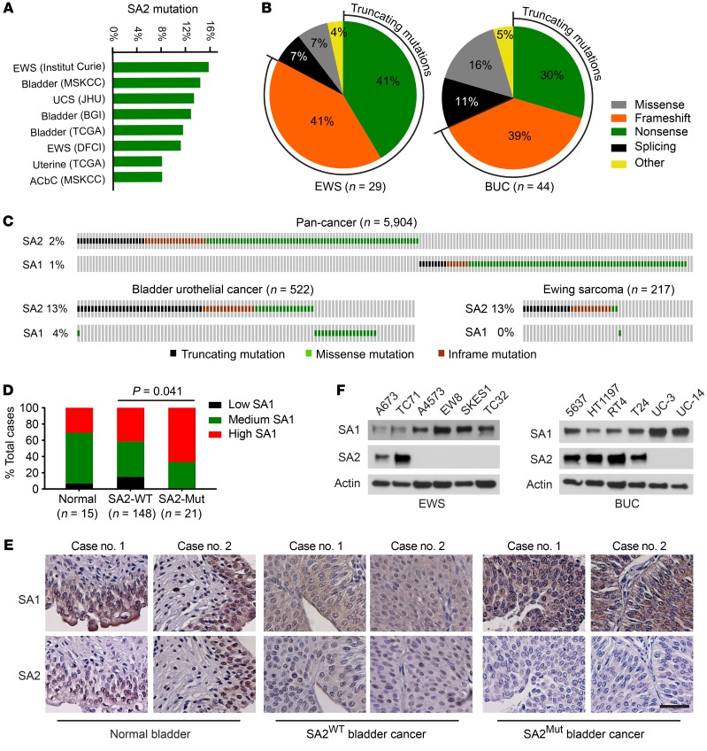
Figure 1. SA2 is frequently mutated in EWS and BUC.
(A) Frequencies of SA2 mutation in a variety of human cancers. UCS, uterine carcinosarcoma; ACbC, adenoid cystic carcinoma of the breast. (B) The nature of SA2 alterations in all EWS (left) and BUC (right) data sets as listed in A. (C) Genomic alterations of SA1 and SA2 in EWS and BUC data sets as listed in A and in 15 other pan-cancer data sets in TCGA. (D and E) Negative correlation between SA1 and SA2 expression levels (D, Fisher’s exact test) and their representative immunohistochemical images (E) in human BUC samples and adjacent normal controls. Scale bars: 50 μm. (F) Protein levels of SA1 and SA2 in human EWS and BUC cell lines, determined by immunoblotting. β-Actin was used as a loading control. Experiments were conducted 3 times for validation.

