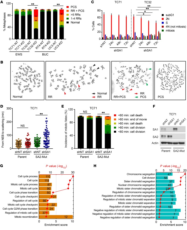
Figure 4. Depletion of SA1 in SA2-mutated cells leads to lethal mitotic retardation and failure.
(A and B) Quantification of cohesion defects (A) and representative metaphase images (B) in EWS and BUC cells expressing Dox-inducible control shNT or SA1-specific shSA1. (C) Cell-cycle analysis of the SA1-depleted TC71 and TC32 cells by costaining with DAPI and phospho–histone H3. (D) Duration of mitosis in parental and isogenic SA2-mutated TC71 cells expressing Dox-induced control shNT or SA1-specific shSA1. Cells were synchronized with double-thymidine block and measured by differential interference contrast (DIC) microscopy time-lapse imaging for 48 hours after release. (E and F) Under each condition as described above, 50 cells were analyzed. Quantification of mitotic fates is shown in E. KD efficiency of shSA1 in TC71 cells is shown in F. (G and H) Negative enrichment of cell-cycle (G) and chromosome segregation (H) gene sets following SA1 KD in the SA2-mutated TC32 cells as determined by GO enrichment analysis. *P < 0.05; **P < 0.01, Fisher’s exact test (A and E) and unpaired 2-tailed t test (C and D). Data are presented as mean ± SD and are representative of 3 independent experiments (A–F).

