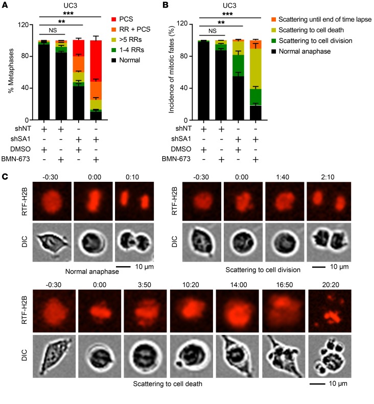
Figure 8. Combined treatment with SA1 depletion and PARP inhibitor in SA2-mutated cells aggravates cohesion defects.
(A) Quantification of cohesion defects in UC3 cells expressing Dox-induced control shRNA or shSA1 with or without BMN-673 (10 nM) treatment. (B, C) Percentages of mitotic fates (B) in UC3 cells after combined treatment with Dox-induced SA1 KD and BMN-673 (10 nM). The representative images for each type of mitotic fate are shown in C. **P < 0.01; ***P < 0.001, Fisher’s exact test. Data are presented as mean ± SD and are representative of 3 independent experiments.

