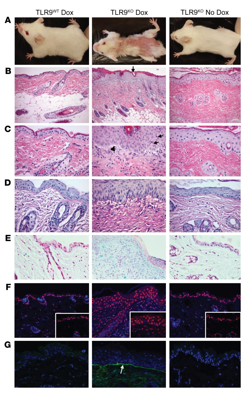
Figure 3. TLR9 deficiency results in LLSI.
(A) Clinical appearance of DO11-injected TLR9WT or TLR9KO Ii-TGO mice, provided or not provided with Dox chow, at 4 weeks after injection. (B and C) H&E-stained skin sections showing follicular plugging (black arrow, B), perivascular and perifollicular mononuclear infiltrate, vacuolization of basal layer (black arrowhead, C), and apoptotic KCs in the epidermis (black arrows, C). Original magnification, ×200 (B); ×400 (C).(D) Basement membrane thickening shown by PAS stain. Original magnification, ×400. (E) Mucin deposition in the dermis detected by Alcian blue stain. Original magnification, ×200. (F) TUNEL stain showing apoptotic cell death (red) counterstained with DAPI (blue). Original magnification, ×200; ×400 (inset). and (G) Ig deposition at basement membrane (white arrow) detected by FITC anti-IgG (green) and counterstained with DAPI (blue). Original magnification, ×200. Images shown are representative of 5 mice per group from 3 independent experiments.

