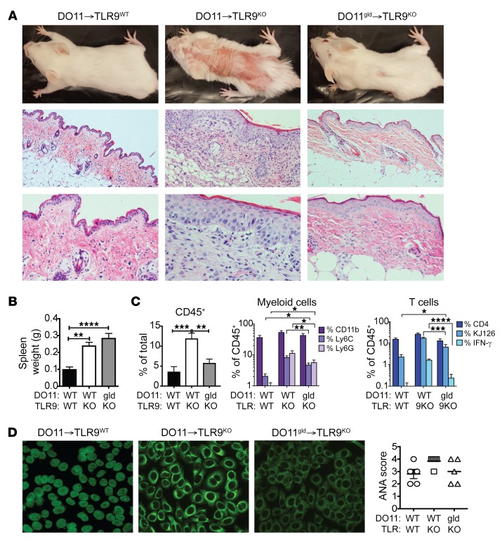
Figure 5. FasL-deficient DO11 T cells fail to induce skin lesions.
DO11 or DO11gld T cell–injected Dox/400R TLR9WT or TLR9KO Ii-TGO recipients were evaluated 4 weeks after injection. (A) Clinical appearance (upper row) and skin histology by H&E staining. Original magnification, ×100 (middle row); ×200 magnification (bottom row). Representative images from n = 5 mice per group. (B) Spleen weights. (C) Skin-infiltrating cells, percentages of CD45+ cells in total skin cell suspension (left); percentages of CD11b+ myeloid cells, CD11b+Ly6C+ inflammatory monocytes, and CD11b+Ly6G+ neutrophils (center); and percentages of CD4+, CD4+KJ126+, and KJ126+IFN-γ+ T cells (right) within the CD45+ gate. (D) Autoantibodies detected by HEp2 staining and ANA score. Original magnification, ×200. Data are shown as mean ± SEM and are representative of 2 independent experiments with n = 8 mice per group. *P < 0.01; **P < 0.01; ***P < 0.001; ****P < 0.0001, 1-way ANOVA and 2-way ANOVA with Šidák’s multiple-comparison test.

