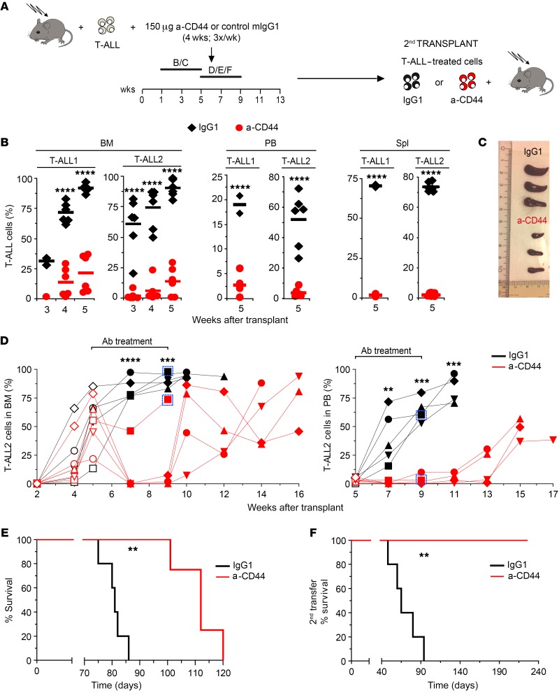
Figure 7. CD44 targeting impairs LIC activity and progression in vivo of human T-ALL and prolongs survival.
(A) RAG-2–/– × γc–/– mice transplanted with primary human T-ALL1 or T-ALL2 cells (Supplemental Figure 4) received 3 weekly i.p. injections of either blocking anti-CD44 mAb (HP2/9) or control IgG1 during 4 weeks, starting at 1 week (B/C) or 5 weeks (D/E/F) after transplant. When indicated, T-ALL cells recovered from the BM of transplanted mice at the end of treatment were transplanted into secondary hosts. (B) Percentages of T-ALL1 and T-ALL2 cells infiltrating the BM, PB, and spleen of mice treated from weeks 1 to 5 after transplant as shown in A. Mean values from 3 independent experiments with a total of 13 to 18 mice for T-ALL and 3 to 8 mice for T-ALL2 are shown. (C) Image of representative spleens obtained at the end of treatment from mice shown in B. (D) Thorough analysis of anti-CD44 in vivo treatment showing percentages of human T-ALL2 cells infiltrating the BM (left) and PB (right) of 5 mice/group treated from weeks 5 to 9 after transplant as shown in A. Empty symbols represent cell percentages before the onset of Ab treatment. Boxes identify individual donors for secondary transplantations shown in F. (E) Kaplan-Meier survival curve of mice treated with anti-CD44 mAb (HP2/9) or control IgG1 in D. (F) Kaplan-Meier survival curve of secondary recipients transplanted with T-ALL2 cells (33 cells/mouse) obtained from the BM of individual donors represented as boxed in D. **P < 0.01; ***P < 0.001; ****P < 0.0001.

