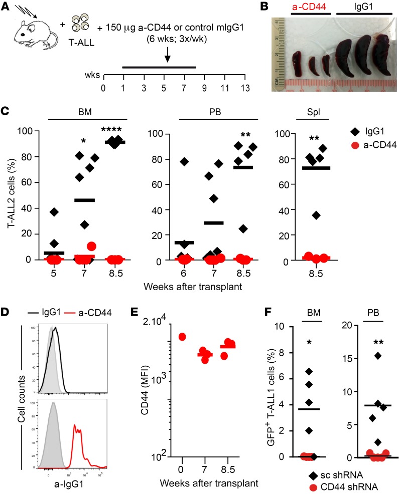
Figure 8. CD44-mediated interaction with the BM microenvironment is required for T-ALL cell leukemogenesis.
(A) NSG mice transplanted with primary T-ALL2 cells received 3 weekly i.p. injections of either blocking anti-CD44 mAb (HP2/9) or control IgG1 during 7 weeks, starting at 1 week after transplant. (B) Image of representative spleens obtained at the end of treatment from mice shown in A. (C) Percentages of T-ALL2 cells infiltrating the BM, PB, and spleen of 5 to 10 mice treated as shown in A. (D) Representative FACS analysis showing persistence of anti-CD44 HP2/9 mAb on the surface of T-ALL cells recovered from the BM of NSG mice represented in A at the end of treatment with either anti-CD44 mAb or IgG1. Bound HP2/9 mAb was detected by reactivity with PE-labeled anti-mouse IgG1 (n = 3). (E) FACS analysis showing levels of HP2/9 anti-CD44 mAb persisting on the surface of T-ALL2 cells that infiltrate the BM of anti-CD44–treated mice shown in A at the indicated weeks after transplant. Shown are MFI values from a total of 7 mice, determined as in D. (F) BM and PB infiltration of T-ALL1 cells transduced with a lentiviral vector encoding GFP and either CD44-specific shRNA (28% transduction efficiency) or a scramble control shRNA (36%). Data show percentages of GFP+ cells within infiltrating CD45+ T-ALL1 cells of 5 NSG mice/group at 11 weeks after transplant. *P < 0.05; **P < 0.01; ****P < 0.0001.

