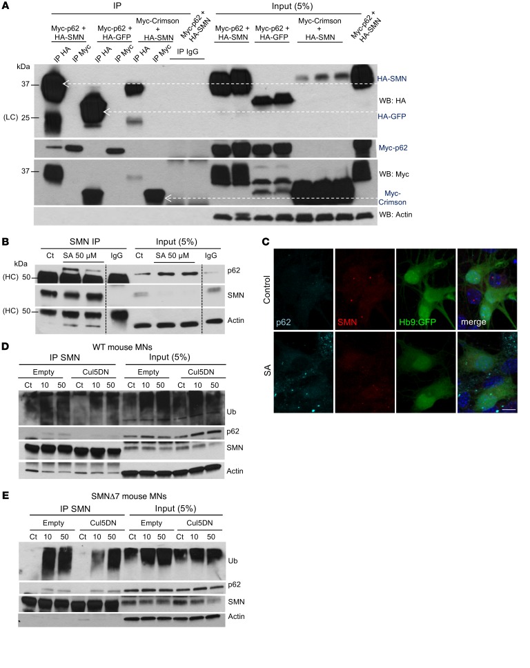
Figure 3. p62 interacts with SMN.
(A) HA and Myc representative IP from HEK293T lysates transfected with the indicated plasmids and immunoblotted against HA, Myc, and p62. WB, Western blot. (B) Representative IP of endogenous SMN protein from mouse SMA MN lysates treated with control or SA for 6 hours and immunoblotted against p62, SMN, LC3, and actin. (C) Representative image from mouse SMA ESC–derived MNs cultured in control conditions or with SA and immunostained against p62 (cyan) and SMN (red) (MNs express Hb9:GFP, and nuclei are stained with DAPI, blue). Scale bar: 10 μm. (D) Representative IP of endogenous SMN protein from WT and SMA mouse MNs. (E) MN lysates infected with lentivirus expressing the DN form of Cul5 for 5 days compared with empty vector–infected cells. MN cultures were treated with control media or SA (10 or 50 μM) for the last 6 hours of the culture. Membranes were immunoblotted against ubiquitin, p62, SMN, and actin. HC, heavy IgG chain; LC, light IgG chain.

