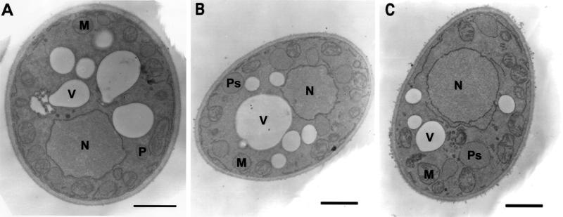
Figure 3.
Ultrastructure of wild-type and of pex19 mutant cells. E122 (A), pex19-1 (B), and pex19KOA (C) strains were grown in glucose-containing YEPD medium for 16 h, transferred to oleic acid–containing YPBO medium, and incubated for an additional 8 h. Cells were fixed in 1.5% KMnO4 and processed for electron microscopy. M, mitochondrion; N, nucleus; P, peroxisome; Ps, peroxisomal structure; V, vacuole. Bar, 1 μm.

