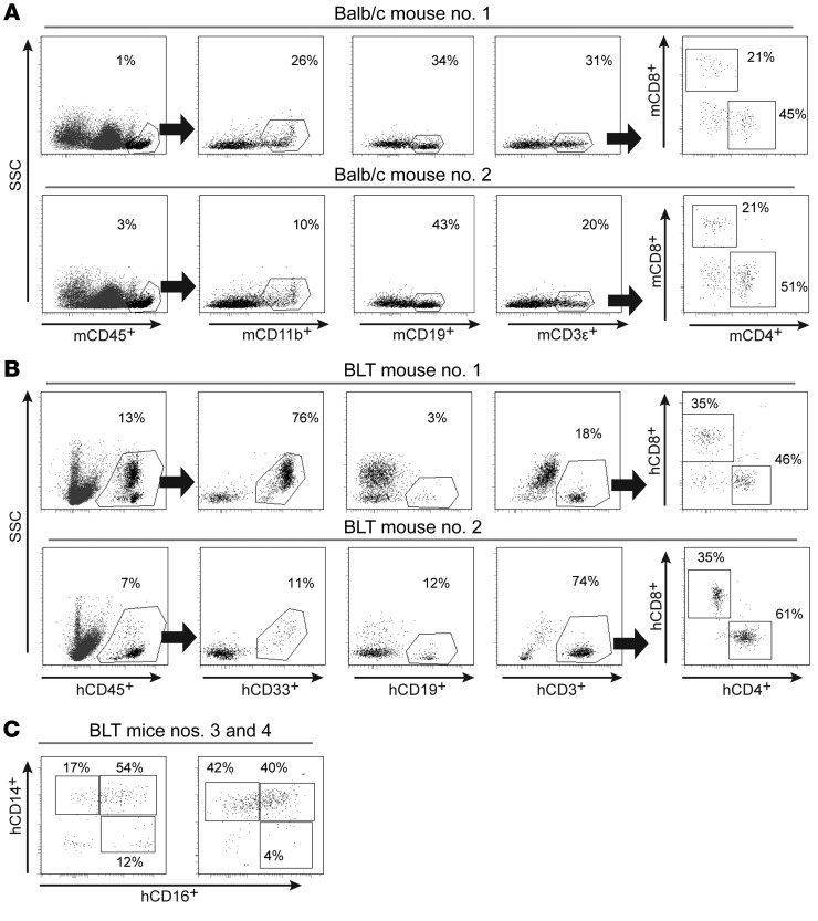
Figure 1. Hematopoietic cells are present in the brains of WT and BLT humanized mice.
(A) Flow cytometric analysis revealed the presence of murine hematopoietic cells (mCD45+) in the brains of BALB/c mice. Murine myeloid cells (mCD11b+), B cells (mCD19+), and T cells (mCD3ε+), including CD4+ and CD8+ T cell subsets, were present. (B) Representative flow cytometric plots from 2 of the BLT mice in Figure 2A demonstrating the presence of human hematopoietic cells (hCD45+), myeloid cells (hCD33+), B cells (hCD19+), and T cells (hCD3+), including CD4+ and CD8+ T cell subsets. (C) Phenotypic characterization of the human macrophages in the brains of BLT mice showed the presence of classical (CD14+CD16–), intermediate (CD14+CD16+), and nonclassical (CD14dimCD16+) macrophages (gating for hCD45+hCD11b+CD33+ cells).

