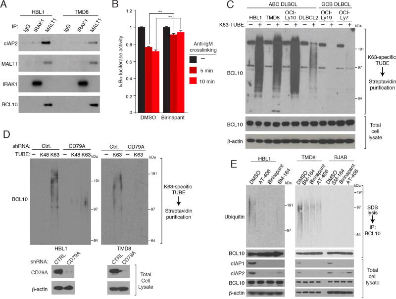
Figure 3. cIAP1/2 participate in chronic active BCR signaling in ABC DLBCL.
(A) Immunoprecipitation (IP) from ABC DLBCL lines using the indicated antibodies were immunoblotted for the indicated proteins. (B) Relative luciferase activity of an IκBα-luciferase fusion protein in TMD8 cells treated with birinapant (5µM) for 6 hours and then with an anti-IgM antibody (10 µg/ml) for the indicated times. Reporter activity was normalized to values from DMSO-treated samples at time zero. Error bars denote SEM of triplicates. p values (Student’s t test) compare treatment groups with the DMSO control; ** p < 0.01. (C) SDS lysates of the indicated lines were subjected to biotin-labeled K63-specific TUBE binding, streptavidin purification. TUBE-purified proteins or total lysates were analyzed by immunoblotting. (D) ABC DLBCL lines were induced to express a CD79A or control (Ctrl.) shRNA. SDS lysates were mixed with biotin-labeled, K63-specific or K48-specific TUBEs followed by streptavidin purification. TUBE-purified proteins or total lysates were analyzed by immunoblotting. (E) SDS (1%) lysates prepared from DLBCL lines treated with the indicated SMAC mimetics (5 µM) for 24 hours, diluted and subjected to anti-BCL10 IP. IPs or total lysates were analyzed by immunoblotting. See also Figure S3.

