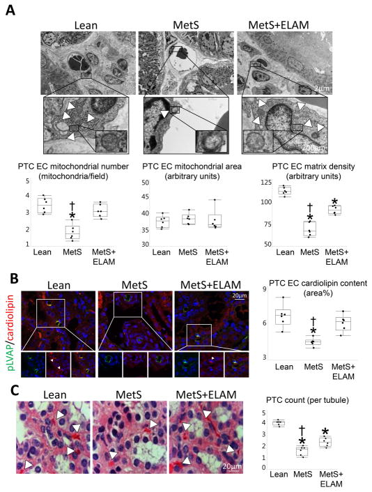
Figure 2. ELAM attenuates endothelial cell (EC) mitochondrial injury and restores cardiolipin content.
A: Transmission electron microscopy and quantification of PTC-EC mitochondrial number and area, and matrix density (5 PTC-ECs per animal, 6 animals/group). B: Representative double immunofluorescent staining (40X) for the PTC-EC marker plasmalemma vesicle associated protein (pLVAP, green) and the mitochondrial inner membrane phospholipid cardiolipin (red) showing decreased endothelial mitochondria and cardiolipin expression (merge yellow) in MetS, which was normalized in ELAM-treated pigs (n=6/group). C: Representative H&E-stained kidney sections (x40 images). The number of capillaries per tubule decreased in MetS compared to Lean, but improved in MetS+ELAM (n=6/group). *p<0.05 vs. Lean, †p<0.05 vs. MetS+ELAM.

