
Figure 3. Expression of viperin in HEK293 cells produces ddhCTP.
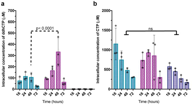
a, Cells expressing Hs viperin (aqua), Hs viperin and Hs CMPK2 (maroon), or empty vector (dark blue). Analysis of ddhCTP formation indicates that the Hs viperin + Hs CMPK2 cells show a statistically significant increase in ddhCTP formation over viperin alone at 48 hr post transfection. In cells with empty vector, ddhCTP levels were undetectable. b, Intracellular concentrations of CTP did not differ significantly (ns) over time (n = 3 biologically independent samples, mean ± S.D., two-way ANOVA, Tukey post-hoc).