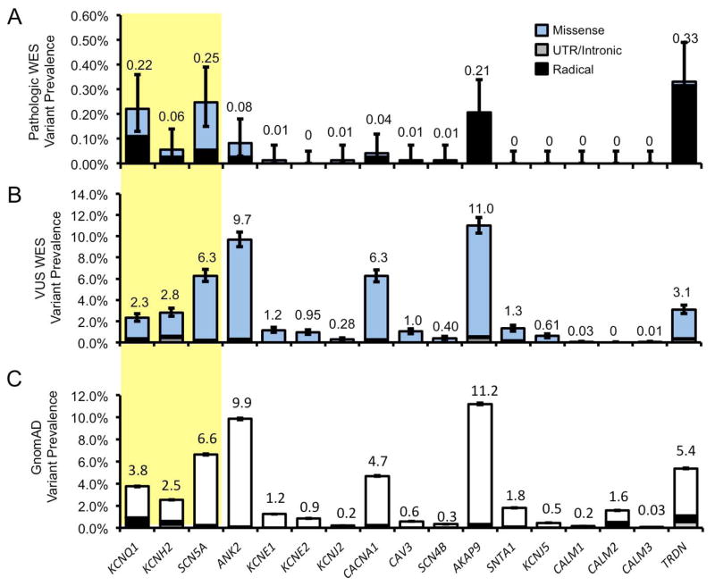
Figure 3.
WES and control cohort gene-specific variant prevalence. A, Bar graph of WES variants (blue fill) deemed “likely pathogenic” at time of genetic testing for each LQTS-associated gene. B, Variants deemed variants of undetermined significance (VUS). C, Rare variants among GnomAD control cohort (white fill). Missense (blue/white), intronic and untranslated regions (UTR, gray), and radical mutations (black) are noted. Error bars denoted 95% CI.

