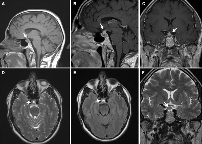
Fig. 1.
Magnetic resonance imaging of the pituitary gland after 2 cycles of ipilimumab obtained in the setting of new headache. Sagittal T1 precontrast (A), sagittal T1 postcontrast (B), and coronal T1 postcontrast (C) images demonstrate a 1.5 cm heterogeneously enhancing mass filling the sella and extending into the suprasellar cistern and up the pituitary infundibulum to abut the undersurface of the midportion of the optic chiasm. Axial T2 (D), axial T2 fluid attenuation inversion recovery (E), and coronal T2 (F) images demonstrate a 7 mm pocket of fluid signal within the sellar portion of the mass, likely representing a small internal subacute hematoma versus other proteinaceous fluid.

