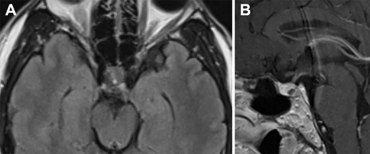
Fig. 2.
One-month follow-up magnetic resonance imaging of the pituitary gland after the ipilimumab was given and corticosteroid therapy was initiated. Axial T2 fluid attenuation inversion recovery (A) and sagittal T1 postcontrast (B) images demonstrate resolution of sellar mass with only a small focus of residual T2 signal abnormality.

