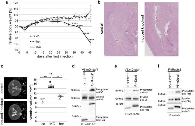
Fig. 5. Ruvbl1 deletion in multiciliated cells results in hydrocephalus in mice.
a Induction of Ruvbl1 knockout (iKO; n = 34) in multiciliated cells by tamoxifen injection (“day 0”) led to significant weight loss compared with control (co; n = 31) or heterozygous (het; n = 8) littermates. Repeated measures two-way ANOVA (F(26,906) = 8.821, p < 0.0001) with Bonferroni-corrected post hoc analyses (****p < 0.0001). b Histological PAS staining of brain tissue showing that the induction of Ruvbl1 knockout in ependymal cells led to widening of brain ventricles (asterisks). Scale bars: 1 mm. c MRI scan of brain showing significantly increased ventricle volumes in the induced knockout mice (iKO; n = 4) compared with control (co; n = 3) or heterozygous (het; n = 3) littermates (F(2, 7) = 34.47, p = 0.0002; Tukey’s post hoc ***p < 0.001). Mean ± SEM. d Dnaaf1 specifically co-precipitated with Ruvbl1, but not with the control protein. FLAG-tagged Ruvbl1 or control protein (EPS) was precipitated, and precipitates were analyzed for co-precipitating V5-tagged Dnaaf1. e Dnaaf1 specifically co-precipitated with Dpcd, but not with a control protein. FLAG-tagged Dpcd or control protein (EPS) was precipitated, and precipitates were analyzed for co-precipitating V5-tagged Dnaaf1. f Ruvbl2 specifically co-precipitated with Dpcd, but not with a control protein. FLAG-tagged Dpcd or control protein (EPS) was precipitated, and precipitates were analyzed for co-precipitating V5-tagged Ruvbl2

