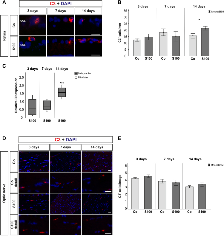
Figure 2.
Mild C3 activation. (A) To evaluate an activation of C3, retinal cross-sections were stained with anti-C3 (red) and DAPI (cell nuclei, blue) 3, 7, and 14 after immunization. (B) No alterations in C3+ cell numbers were noted in the S100 group after 3 and 7 days (p > 0.05). After 14 days, significantly more C3 depositions were observed (p = 0.04). (C) The C3 mRNA expression level showed no alterations 3 and 7 days after immunization (p > 0.05). A significant higher level of C3 mRNA was observed at 14 days (p < 0.001). (D) Anti-C3 staining (red) of the optic nerves was performed 3, 7, and 14 days after immunization. Cell nuclei were labeled with DAPI (blue). (E) No changes were revealed at all points in time (p > 0.05). Abbreviation: GCL = ganglion cell layer. Values are mean ± SEM for immunohistology and median ± quartile + maximum/minimum for qRT-PCR. Scale bars: 20 µm.

