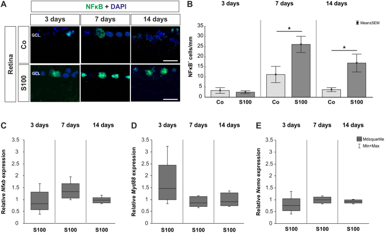
Figure 4.
Increased NFκB staining. (A) Retinas were labeled with an anti-NFκB antibody (green) and DAPI (blue). (B) At 3 days, no changes were observed in regard of the number of NFκB+ cells in the S100 group (p > 0.05). After 7 days, significantly more NFκB+ cells were noted in S100 animals (p = 0.03). A significant increase of NFκB+ cells was still revealed in this group after 14 days (p = 0.03). (C) QRT-PCR analysis revealed no differences in Nfκb mRNA expression at all points in time (p > 0.05). However, a slight trend towards an upregulation could be observed at 7 days (p = 0.18). (D) The expression levels of Myd88 showed a trend towards an upregulation at 3 days (p = 0.1). At 7 and 14 days, no alterations were noted (p > 0.05). (E) At all points in time, the expression of NEMO mRNA remained unchanged (p > 0.05). Abbreviation: GCL = ganglion cell layer. Values are mean ± SEM for immunohistology and median ± quartile + maximum/minimum for qRT-PCR. Scale bars: 20 µm.

