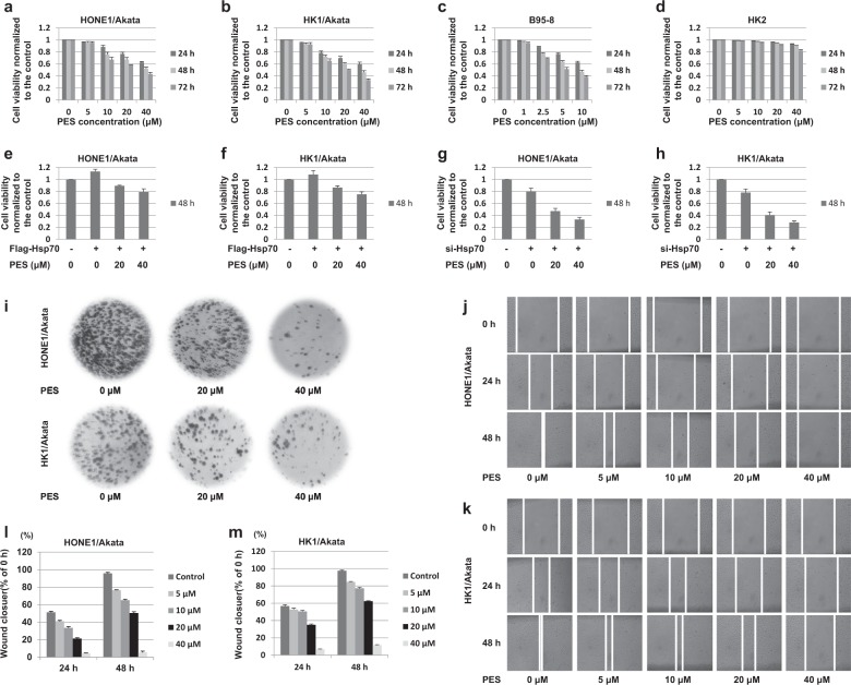
Fig. 3. PES inhibits proliferation and migration of EBV-positive cells.
a HONE1/Akata, b HK1/Akata, c B95-8 and d HK2 cells were treated or not with increasing concentrations of PES for 24, 48, and 72 h, respectively. e HONE1/Akata and f HK1/Akata cells were transfected with the pEF-Flag-Hsp70, followed by treatment with PES (20, 40 μM) beginning at 4 h after transfection. g HONE1/Akata and h HK1/Akata cells were transfected with Hsp70 siRNA, followed by treatment with PES (20, 40 μM) beginning at 4 h after transfection. Cell viability was determined by CCK-8 assays. i HONE1/Akata and HK1/Akata cells were treated or not with PES (20 or 40 μM) and cultured for another 15 days. Colony formation assay was carried out to determine the long-term effect of PES on cell proliferation. HONE1/Akata (j,l) and HK1/Akata (k,m) cells were treated with the linear scratch wounds, followed by the treatment of PES (0, 5, 10, 20 or 40 μM) for 24 and 48 h. Then the migration of the cells towards the wound was visualized. Photographs were captured and analyzed using Image J software

