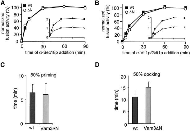
Figure 6.
Vam3ΔN vacuoles show a delay in docking. Standard fusion reactions (200 μl), containing wild-type or Vam3ΔN vacuoles, were incubated in the presence of cytosol and ATP; at the indicated time points a 30-μl aliquot was mixed with an inhibitor and incubation at 26°C was continued for a total of 90 min. Concentration of antibodies to Sec18p and Vti1p IgGs was 0.1 μg/μl, Gdi1p was used at 64 μg/ml. (A) Sensitivity to α-Sec18p of wild-type and Vam3ΔN vacuoles. Priming kinetics obtained were normalized by setting fusion at 90 min to 100%. An inlet shows the alkaline phosphatase units before normalization. Average values of nine independent experiments are shown. (B) Sensitivity to α-Vti1p or Gdi1p of wild-type and Vam3ΔN vacuoles. Average values of 20 independent experiments are shown. (C) Time point, where fusion inhibition by α-Sec18 is 50%. Wild-type 6.5 ± 1.7 min (SEM), n = 9; Vam3ΔN 6.2 ± 2.2 min (SEM), n = 9. Difference not significant (Student's t test = 0.4). (D) Time point, where fusion inhibition by α-Vti1 or Gdi1p is 50%. Wild-type 11.5 ± 2.9 min (SEM), n = 20; Vam3ΔN 15.3 ± 2.4 min (SEM), n = 20. Difference is highly significant (Student's t test < 0.00001).

