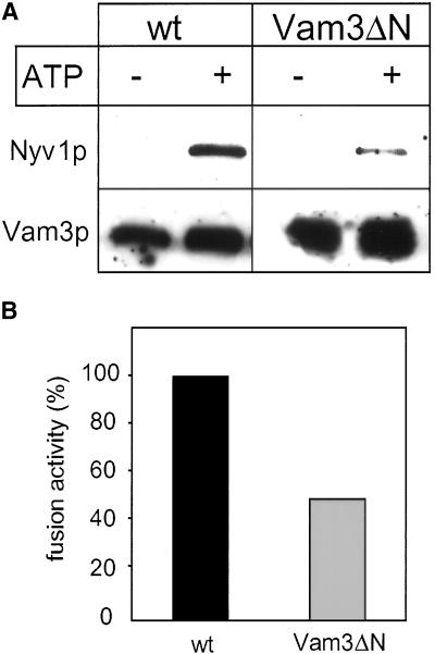
Figure 7.
Vam3ΔN vacuoles form less trans-SNARE complexes. (A) Coimmunoprecipitation of Nyv1p with anti-Vam3p. Vacuoles from BJ3505 nyv1Δ or BJ3505 nyv1Δ VAM3ΔN were fused with DKY6281 vam3Δ as described in MATERIALS AND METHODS. After the incubation vacuoles were collected by centrifugation, solubilized and immunoprecipitated with α-Vam3p. Eluted proteins were separated by SDS-PAGE and detected by Western blotting with antibodies to Nyv1p and Vam3p. The amount of coprecipitated SNAREs was quantified densitometrically (NIH image 1.6): wild-type, 100 ± 10; Vam3ΔN, 34 ± 16 (mean density after background subtraction in arbitrary units ± SEM, n = 3). (B) A 30 μl aliquot was removed from the identical fusion reaction described in (A) and incubated for 90 min at 26°C to measure fusion activity. Fusion of BJ3505 nyv1Δ with DKY6281 vam3Δ (black bar) was set to 100%.

