
Fig. 1.
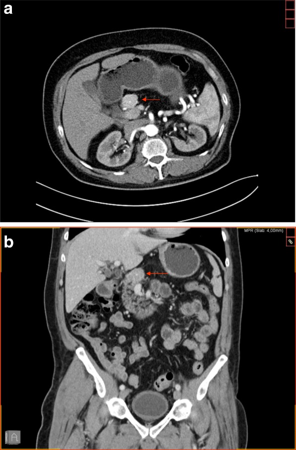
Contrast-enhanced computed tomography scan with axial image in arterial phase (a) and coronal reconstructed image in venous phase (b) showing a round hypervascular mass, measuring approximately 3 cm at the neck of the pancreas with absence of the body and tail of the pancreas. The arrows show the lesion in the pancreas