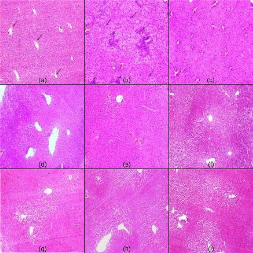
Figure 1.

Representative light micrographs from: (a) normal control showing normal liver histology with radial arrangement of hepatocyte rays around central veins (arrows) that are separated by sinusoids; (b) DEN-treated mice showing HCC of grade (3) with disturbed architecture and loss of lobular pattern, hepatocellular atypia, pleomorphism, intralobular inflammatory infiltrate (arrowheads), malignant hepatocytes with moderate to marked nuclear anaplasia (arrows). Moreover, intralobular inflammatory infiltrates, fibrous tissue deposition (arrowheads) and congested blood vessels are evident. Micrographs from mice treated with sorafenib, (c), perindopril (d), fosinopril (e), losartan (f), perindopril plus sorafenib (g), fosinopril plus sorafenib (h), and losartan plus sorafenib (i) showing regression of malignant changes with almost restoration of lobular architecture and lowering of the grade of HCC to grade 1 (H&E x100)
