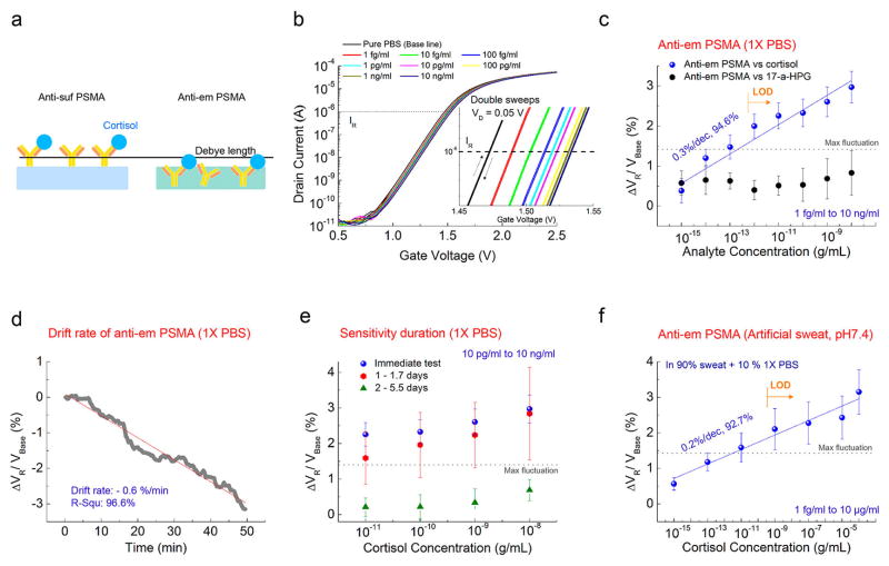
Figure 3.
(a) Presumed schematic image of antibody-embedded geometric in the PSMA polymer matrix. (b) Representative transfer curves of antiem PSMA versus cortisol concentration in 1× PBS. Inset: Close-up transfer curves. (c) ΔVR response of antiem PSMA in terms of the cortisol and 17-α-HPG concentration VR in a 1× PBS solution. ΔVR increases by 0.3% with increasing cortisol over eight samples of antiem PSMA. Random signal in terms of 17-α-HPG over four samples of antiem PSMA. (d) Drift properties of antiem PSMA in 1× PBS over 50 min after stabilization. A negative slope was obtained like that of Figure 2d. (e) Cortisol sensitivity of antiem PSMA as a function of days stored, in a range of cortisol from 10 pg/mL to 10 ng/mL (10 and 6 samples for immediate and stored samples, respectively). Random signals appeared for six samples 2 days after storage. (f) Cortisol sensitivity of antiem PSMA in artificial sweat with pH 7.4 in a range from 1 fg/mL to 100 μg/mL for five samples. 10% 1× PBS was added to the artificial sweat. Three other samples gave little or no response.

