
Fig. 4.
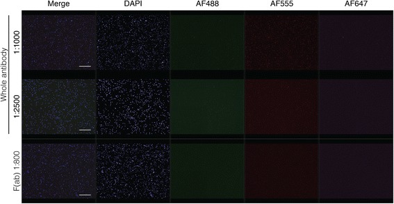
Optimization of secondary antibody application in human bone marrow. A) Comparison of different dilutions of whole secondary antibody and F(ab) fragment secondary antibody in primary unstained bone marrow. Goat anti-rabbit secondary antibodies for all three fluorophores were applied. Scale bar = 50 μm